Why is the Effective Dose Still the Most Critical Parameter in Nuclear Medicine?
Effective Dose: Not Just a Number, but a Vital Indicator
The content shared here is a summary of the article, for the full article: visit health physics
Effective dose is a conceptual measure that evaluates the biological effect of ionizing radiation on the human body.
Simply put, this value refers to which organ is exposed to how much radiation and how this energy is reflected in the risk of developing cancer .
The International Commission on Radiation Protection (ICRP) determines the tissue weighting factors used in these calculations.
For example, organs such as the lungs, bone marrow or thyroid are considered more sensitive to radiation; The contribution of tissues such as skin or muscle is lower.
For a nuclear medicine specialist, the importance of the effective dose is twofold:
- To show whether the total amount of radiation received by the patient is within safe limits,
- To compare the dose efficiency of different imaging protocols.
For this reason, “effective dose” is still used as the main criterion of radiation safety policies today.
The Role of Radiopharmaceuticals: An Anatomical Map of Internal Radiation
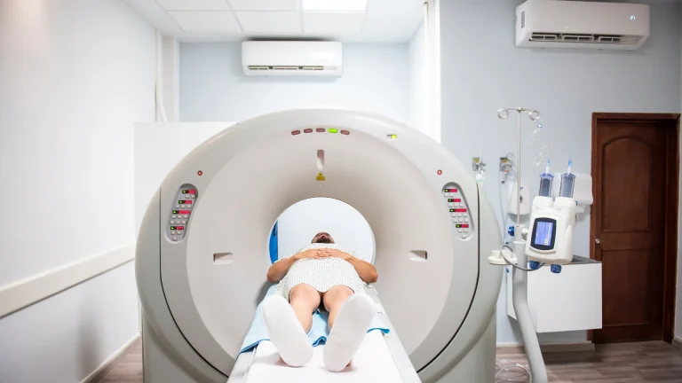
The most special aspect of nuclear medicine is that the radiation comes from inside the body.
A radiopharmaceutical is injected into the patient and follows different biokinetic pathways according to its involvement in organs.
The two key substances used in this process — 18F-FDG (glucose tracer) and 68Ga-PSMA (prostate-specific membrane antigen) — accumulate in different organs throughout the body.
While 18F-FDG releases high energy, especially in the heart and bladder wall, it has been determined that 68Ga-PSMA is mostly retained in the kidneys.
According to the results of the research:
- The mean total effective dose ≈ in 18F-FDG PET/CT scans was 25.3 mSv,
- In 68Ga-PSMA PET/CT scans, the ≈ was calculated as 22.0 mSv .
These values indicate that the CT portion contributes much more to the dose than the PET component — the CT contribution is 75% in 18F-FDG and 92% in 68Ga-PSMA.
The Difference Between the Software: IDAC-Dose 2.1 vs. OLINDA
So why do different results occur when the data of the same patient is used?
The answer to this is hidden in the software’s approach to biokinetic modeling and phantom geometry .
OLINDA/EXM is built on the MIRD methodology that has been used since the 1980s.
This system uses simplified geometric representations of the human body, called “mathematical (stylized) phantoms“.
Therefore, energy transfer between organs is estimated by a more general approach.
In contrast, IDAC-Dose 2.1 uses the voxel phantom models developed by ICRP 110 — i.e., three-dimensional anatomical models created from real human CT data.
This approach represents organ volumes and tissue transitions much more accurately.
As a result, the PET/CT data of the same patient produced quite different effective dose results in the two software.
The following table summarizes these differences
Comparison of IDAC-Dose 2.1 and OLINDA/EXM Software
| Feature / Parameter | OLINDA/EXM | IDAC-Dose 2.1 |
|---|---|---|
| Basic Methodology | MIRD (Medical Internal Radiation Dose) | Advanced modeling based on ICRP 128 and 110 guidelines |
| Phantom Type | Stylized phantom | Voxel (3D real human anatomy) phantom |
| Data Source | Standard reference values (ICRP 60, 1990) | Real CT data (ICRP 103, 2007) |
| Tissue Weight Factor | ICRP 60 | ICRP 103 |
| Radiopharmaceutical Modeling | Simplified biokinetic data | Updated biokinetic dataset |
| Effective Dose (18F-FDG, PET) | 9.96 mSv | 6.28 mSv |
| Proximity to Clinical Reality | Intermediate – estimated | High – individualized modeling |
| Advantage | Easy-to-use, classic model | More accurate organ and tissue representation |
| Disadvantage | Old tissue coefficients, tendency to overdose estimation | More complex data entry and processing time |
This difference clearly shows that the calculation of the effective dose in nuclear medicine depends not only on the amount of activity injected but also on the geometry of the mathematical model used.
In other words, when the biokinetic model used for the same patient changes, the radiation safety assessment may also change.
Therefore, in modern clinical practice, the use of voxel-based software such as IDAC-Dose 2.1 is now a necessity for both scientific accuracy and patient safety .
ICRP 60 and 103: Generational Difference in Radiation Risk
Radiation safety guidelines have also evolved over time.
In the ICRP 60 (1990) model, tissue weight factors were determined based on limited clinical data.
The newly published ICRP 103 (2007) standard re-evaluated organ sensitivities with long-term epidemiological studies.
For example, the risk coefficient of breast and thyroid tissues is increased; the liver is reduced. These changes have enabled new software, such as IDAC-Dose 2.1, to produce lower and more realistic effective doses .
In other words, as science has advanced, the meaning of measurement , not measurement, has changed. This, in turn, helps protect patients from unnecessary high-risk predictions.
Clinical Reality: 90% of the Effective Dose Originates from CT
One of the most striking findings in the study is the predominance of CT’s contribution to the total effective dose.
In other words, the patient receives more radiation due to the X-rays he is exposed to in the tomography (CT) part than the radiopharmaceutical used for PET.
In the study, the average CT effective doses were calculated as follows:
- 18F-FDG protocol: 19.05 ± 5.89 mSv
- 68Ga-PSMA protocol: 20.17 ± 5.87 mSv
These differences highlight the importance of low-dose CT protocols.
Modern hospitals now use dose monitoring systems to instantly monitor the total radiation received by each patient. The goal is simple: to adhere to the principle of “lowest reasonably achievable dose” (ALARA).
The Human Side of the Story: Through the Eyes of an Expert
For a nuclear medicine specialist, an “effective dose” is not just a statistic; It is a decision that he makes hundreds of times in each scan.
When a patient enters the PET/CT device, the physician has to rely on the software in hand.
If the calculation model is outdated, the patient may receive more radiation than necessary — or the tumor may be misjudged.
The goal is to achieve “individualized dose optimization” tailored to each patient.
In the near future, these calculations are expected to be automated with artificial intelligence-supported systems .
Machine learning algorithms have the potential to perform instant radiopharmaceutical dose optimization based on patient age, gender, metabolic activity and organ volumes.
Looking to the Future: Between Science, Ethics, and Technology
The reason why the effective dose is still the most critical parameter in nuclear medicine is that it carries both a scientific and ethical responsibility .
More than every millisievert, it’s about the safety of a patient.
The PET/CT technologies of the future promise not only higher resolution images, but also systems that operate with lower risk .
Dose planning software integrated with artificial intelligence will be able to analyze individual biokinetic data based on the ICRP 103 guideline.
After all, the power of unseen light will no longer be used not only for diagnosis, but also to protect human health .
So, do you think the nuclear medicine of the future will be able to achieve a “zero risk” imaging protocol specific to each patient?